
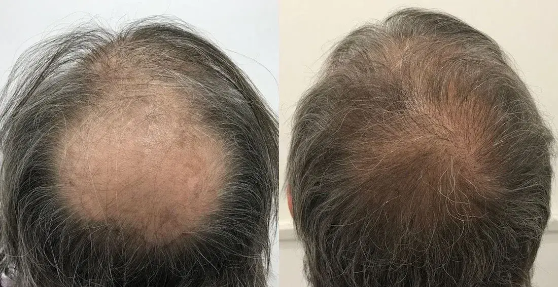
Crown hair transplants do not automatically fail, but a result may appear unsuccessful because of poor graft growth, insufficient coverage, continuing loss of native hair, an unnatural whorl design, or evaluation before development is complete. The crown’s size and spiral growth pattern can also make satisfactory graft growth look less dense than expected. A proper assessment must identify which problem is present.
The crown, also called the vertex, is the circular or oval area toward the upper back of the scalp. Several different outcomes can affect this region, and they should not all be grouped under the label “failed crown hair transplant.”
Does a Slow Crown Result Mean the Transplant Has Failed?
No. An incomplete-looking crown result does not automatically mean that the grafts failed.
Transplanted hairs may shed after surgery before new shafts become visible. Growth then develops gradually, and different follicles may become noticeable at different times. The International Society of Hair Restoration Surgery states that a full result may take a year or longer to appear in some patients.
Slow development must be distinguished from:
Temporary postoperative shedding: Hair shafts fall while follicles remain in the scalp.
Uneven early development: Some areas become visible before others.
Low visual density: Hair grows, but the scalp remains visible.
Persistent poor growth: Large areas show little or no development after an appropriate observation period.
Progressive native hair loss: Transplanted hair grows, but untreated surrounding hair becomes thinner.
There is limited high-quality evidence proving that crown follicles always grow more slowly for biological reasons. In practice, the crown can appear slower because it covers a broad area, hair points in several directions, and overhead light exposes more scalp. The procedure date, photographs, scalp findings, hair characteristics, and treatment plan should guide the evaluation rather than one universal deadline.
Why Is the Crown More Difficult to Restore?
The crown presents both technical and visual challenges.
The whorl changes direction continuously
Natural crown hair usually rotates around one or more central points. Grafts must follow changing angles and directions rather than a single straight pattern. Research examining crown follicle orientation also shows that natural follicular-unit orientation is more complex than simply drawing a spiral on the scalp.
Incorrect direction may produce crossed hairs, separated sectors, or a visibly broken whorl even when grafts grow.
The treatment area can be large
A crown may require coverage across a broad circular area. Spreading a limited number of follicular units across that surface can produce lower visual density than concentrating the same donor supply in a smaller frontal zone.
Light reaches the scalp from above
The crown faces overhead lighting directly. Hair also separates around the whorl center, making scalp visibility more noticeable. Hair shaft caliber, curl, color contrast, length, and styling all affect the visual result.
Donor hair is limited
Hair transplantation redistributes existing follicles; it does not create an unlimited new supply. Donor assessment and graft allocation therefore affect how much coverage can reasonably be achieved. The donor area remains one of the principal limits of transplantation, regardless of the extraction or implantation method.
Future hair loss must be considered
Androgenetic alopecia is progressive. Native hair around a transplanted crown can continue to miniaturize, eventually leaving a ring, gap, or enlarged area around previously transplanted follicles.
Crown planning must therefore consider the frontal scalp, mid-scalp, future loss pattern, patient age, and lifetime donor reserve—not only the bald area visible on the day of surgery.
Main Reasons a Crown Hair Transplant May Produce a Poor Result
The Result Was Evaluated Too Early
Primary category: Recovery and expectations
New growth does not appear immediately after transplantation. Early shedding, temporary redness, short hairs, and uneven development can all occur during recovery.
A crown may look especially sparse during this period because new hairs remain short and provide little overlap. As the shafts lengthen and mature, they may create more coverage. Hair volume is influenced by shaft width and length as well as the number of visible hairs.
A meaningful review should compare standardized photographs and clinical findings rather than rely on a single mirror view or different lighting conditions.
Too Few Grafts Were Available for the Treatment Area
Primary category: Planning and donor limitations
The crown may remain visibly thin when the treatment surface is too large for the available donor supply. This does not always mean that the implanted follicles failed to grow.
A responsible plan may intentionally prioritize coverage over high density or preserve donor hair for the frontal and mid-scalp regions. Conversely, a plan that promises dense crown restoration without accounting for future loss may use too much of a limited donor reserve.
Quantitative donor assessment should consider follicular-unit density, hair quality, miniaturization, safe donor boundaries, and likely future requirements. Professional FUE guidance specifically recommends assessing donor follicular-unit density and hair quality before deciding how much can safely be removed.
Grafts Were Distributed Poorly
Primary category: Planning and surgery
The visible result depends on where grafts are concentrated, how the transition zones are built, and how the whorl center connects with the surrounding scalp.
A poorly distributed transplant may have:
Unnecessary density in one small sector.
Wide gaps elsewhere.
An isolated central patch.
Abrupt transitions into thinning native hair.
Too little support around the edge of the crown.
An allocation that ignores likely future expansion.
Evenly spacing grafts does not always create the most natural visual result. Distribution should respond to the existing whorl, hair caliber, scalp contrast, crown size, surrounding hair, and realistic coverage priorities.
The Natural Crown Whorl Was Not Recreated Correctly
Primary category: Design and surgery
A whorl-design error occurs when recipient sites do not follow the patient’s natural rotational pattern. The result may look unnatural even if most grafts survive.
Possible problems include:
A center placed in an unsuitable position.
Hair directed against the remaining native pattern.
Sudden changes in rotation.
Several disconnected directional zones.
Grafts inserted too upright.
A spiral that does not blend with surrounding hair.
Hair transplant practice guidance describes vertex reimplantation as challenging, and specialist hair restoration education treats crown-whorl reconstruction as a technically demanding design task.
Hair Loss Continued Around the Transplanted Grafts
Primary category: Biology and long-term planning
Transplantation may move follicles that are less susceptible to androgenetic miniaturization, but it does not automatically stop loss in untreated native hair.
A result can therefore look satisfactory initially and deteriorate later as the crown expands around it. The transplanted hairs may still be present while the surrounding native hairs become finer or disappear.
The ISHRS identifies continued genetic hair loss around transplanted hair as a common reason patients later seek corrective work.
Medical management may be considered for suitable patients after an individual clinical assessment. Medication is not appropriate for everyone and should not be started, stopped, or changed solely because of general online advice.
The Donor Area Was Weak or Poorly Assessed
Primary category: Candidate selection and planning
A donor area may be unsuitable when it has low density, fine hair, diffuse miniaturization, scarring, previous overharvesting, or follicles taken from regions at risk of future loss.
Weak donor hair can reduce coverage even when it grows. Fine, straight hair with strong hair-to-skin contrast may expose more scalp than thicker or curlier hair.
Extracting beyond a reasonably safe donor zone may also create future loss of transplanted or remaining donor hair. FUE practice guidance warns that grafts removed from outside safer donor areas may be vulnerable to later loss, particularly in younger patients with ongoing alopecia.
Grafts Were Damaged During Extraction, Storage or Implantation
Primary category: Surgery and graft handling
Follicular units can experience mechanical and metabolic stress during extraction, preparation, storage, and implantation.
Potential factors include:
Transection during extraction.
Crushing or drying.
Excessive handling.
Unsuitable storage conditions.
Prolonged time outside the body.
Trauma during loading or implantation.
Placing a graft too deeply or too superficially.
A review of graft survival identifies dehydration, temperature, ischemia, mechanical injury, and handling as factors that can influence growth. Preservation solutions can also affect follicular viability, although clinical outcomes depend on the full process rather than one variable alone.
Because the grafts come from the patient, classic immune “graft rejection” is not usually the first explanation for poor growth. The assessment should instead examine surgical handling, scalp biology, healing, disease, and the treatment plan.
Recipient-Site Trauma, Scarring or Complications Affected Growth
Primary category: Surgery, healing, and biology
The recipient area must support healing around newly placed follicular units. Excessive trauma, dense incision patterns, pre-existing scar tissue, vascular injury, infection, necrosis, or persistent inflammation may affect growth.
Recognized complications of hair transplantation include infection, scarring, folliculitis, persistent redness, tissue necrosis, and unnatural results, although severe complications are not expected in routine uncomplicated recovery.
Scarring does not automatically prove that all grafts failed. A specialist must examine its extent, activity, texture, blood supply, and relationship to the empty areas.
An Underlying Scalp or Hair-Loss Condition Was Not Identified
Primary category: Diagnosis and biology
Not every crown-thinning pattern results from ordinary androgenetic alopecia. Alopecia areata, diffuse unpatterned hair loss, inflammatory scalp disease, or a scarring alopecia may alter candidacy and expected growth.
Subtle lichen planopilaris can overlap with pattern hair loss and may require trichoscopy, clinical assessment, or biopsy when suspected. Poor growth accompanied by persistent redness or perifollicular scaling may require investigation rather than automatic revision grafting.
Central centrifugal cicatricial alopecia can also begin in the crown and may cause itching, pain, scale, bumps, or progressive scarring. These findings are not diagnostic by themselves, but they justify assessment by an appropriately qualified doctor.
Postoperative Trauma or Incorrect Care Affected Early Healing
Primary category: Recovery
Rubbing, scratching, picking crusts, unapproved products, or trauma soon after surgery may disturb healing. However, a poor outcome should not automatically be blamed on the patient.
Postoperative instructions vary according to the procedure, surgeon, and clinical circumstances. Patients should follow the instructions supplied by their treating team rather than replacing them with a generic online schedule.
A review should consider whether a specific event occurred, when it occurred, and whether the grafts or only the visible hair shafts were affected.
The Patient Expected Frontal-Scalp Density in the Crown
Primary category: Expectations and planning
A crown often requires more surface coverage and multidirectional placement than a small frontal area. The same number of grafts may therefore produce less apparent density.
A hair transplant generally creates an impression of coverage rather than restoring the original number of follicles present before hair loss. Some scalp visibility may remain, especially under strong light, with wet hair, or at the center of the whorl.
Expectations should be based on crown-specific cases with comparable crown size, hair caliber, curl, color contrast, styling, donor supply, and degree of native hair loss.
Slow Crown Growth vs Possible Transplant Failure
This table supports discussion with a professional. It cannot diagnose graft failure or a scalp condition.
Observation | May occur during normal development | May justify professional assessment | Why it matters |
|---|---|---|---|
Uneven early growth | Yes | If it remains marked beyond the expected review stage | Follicles do not always become visible simultaneously |
Temporary shedding | Yes | If accompanied by worsening inflammation, scarring, or other symptoms | The shaft may shed while the follicle remains |
Persistently empty areas | Less likely at a later stage | Yes | May reflect poor growth, distribution gaps, scarring, or disease |
Mild early redness | Can occur | If it increases, persists, or returns | Persistent inflammation needs clinical evaluation |
Increasing pain or discharge | No | Seek earlier medical contact | May indicate infection or another complication |
Visible scarring | Some small scars can follow surgery | Yes, particularly if raised, depressed, painful, expanding, or inflamed | Scar tissue can affect appearance and revision planning |
Correct growth but low visual density | Yes | Yes, if substantially different from the agreed plan | May reflect limited graft numbers, fine hair, large surface area, or distribution |
Loss around the transplanted area | Not part of graft maturation | Yes | Native androgen-sensitive hair may continue to miniaturize |
An unnatural or broken whorl | No | Routine specialist review | Suggests a direction or design issue rather than delayed growth |
Patchy crusting, pustules, or worsening tenderness | No | Earlier medical assessment | These can occur with postoperative complications but require examination |
Increasing pain, purulent discharge, worsening redness, tenderness, swelling, or pustules warrant prompt contact with the treating clinic or another qualified medical professional. These signs can occur with infection or inflammatory complications and should not be diagnosed from photographs alone.
When Can a Crown Hair Transplant Be Evaluated Fairly?
There is no single month when every crown result becomes final. Use the following stages as a general framework, not a guaranteed schedule.
Early healing: first days and weeks
The scalp may show redness, swelling, crusting, short implanted shafts, and temporary sensitivity. Some transplanted shafts later shed. At this stage, scalp appearance does not reliably show the eventual density.
Unexpectedly increasing pain, discharge, marked swelling, darkening tissue, spreading redness, or systemic illness requires earlier medical advice.
Early development: first few months
The crown may look similar to its preoperative appearance after shedding. Initial new growth may be fine, short, uneven, or difficult to see under overhead lighting.
A lack of cosmetically meaningful density during this phase does not by itself prove failure.
Intermediate development: several months onward
More follicles may become visible, while existing shafts lengthen and gain cosmetic volume. Standardized photographs become increasingly useful.
At this stage, clinicians can begin assessing distribution, direction, emerging gaps, native-hair loss, and whether development is progressing.
Later assessment: around a year and sometimes longer
Many results can be assessed more fairly around the later part of the first postoperative year, but some patients require longer observation. ISHRS patient guidance notes that full development can take a year or more and that the rate of growth varies between patients.
A final assessment should consider:
Procedure date.
Hair cycle variation.
Standardized photographs.
Hair length and styling.
Shaft caliber.
Crown size.
Graft distribution.
Scalp condition.
Continuing native hair loss.
Any postoperative complications.
Whether further planned sessions formed part of the original strategy.
Signs That the Issue May Be More Than Delayed Growth
Routine review may be appropriate when:
The crown remains thinner than expected.
Growth appears uneven.
The visual density does not match the initial plan.
Native hair around the crown is becoming finer.
The whorl looks unnatural.
The patient cannot determine whether change is occurring.
Earlier medical contact may be appropriate for:
Increasing pain or tenderness.
Discharge or pustules.
Worsening redness or swelling.
Open wounds or darkened skin.
Persistent crusting or inflammation.
Rapidly expanding hair loss.
New smooth, shiny, or visibly scarred areas.
Systemic symptoms such as fever.
Persistently absent growth, clearly defined empty patches, substantial scarring, major directional errors, or a result that remains very different from the agreed plan may warrant a detailed later-stage assessment. None of these findings should be used to self-diagnose infection, necrosis, scarring alopecia, or complete graft failure.
Can a Failed Crown Hair Transplant Be Repaired?
Some crown results can be improved, but not every case is suitable for another operation.
Possible approaches include:
Observation and reassessment
More time may be appropriate when development is still progressing or when standardized photographs do not yet support a reliable conclusion.
Management of continuing native hair loss
A qualified doctor may discuss medical treatment when clinically suitable. The purpose may be to preserve susceptible native hair rather than revive transplanted follicles that did not survive.
No medication is suitable for every patient, and potential benefits, contraindications, side effects, reproductive considerations, and ongoing-use requirements require individual discussion.
Revision hair transplantation
Additional follicular units may be used to improve coverage, fill defined gaps, soften transitions, or reconstruct the whorl. Revision may require one or more stages.
Redistribution or removal of existing grafts
Poorly directed grafts may sometimes be removed, relocated, or visually integrated with additional grafts. The appropriate method depends on their position, direction, scarring, and available donor supply.
Camouflage
Hair fibers, styling changes, scalp concealers, or carefully selected micropigmentation may improve the appearance of low density. ISHRS guidance describes scalp micropigmentation as one option for low-density or scarred regions, including cases with limited donor hair.
Combined approaches
Some patients may need staged surgery, treatment of scalp disease, management of native hair loss, and camouflage rather than one isolated intervention.
Repair suitability depends on:
Remaining donor capacity.
Previous overharvesting.
Crown surface area.
Hair caliber, curl, and color contrast.
Existing graft direction.
Scar tissue.
Scalp health.
Cause of the poor result.
Future loss pattern.
Frontal and mid-scalp priorities.
Realistic density expectations.
The ISHRS notes that limited donor hair can make correction difficult or impossible and that some patients should not undergo further surgery.
PRP, medication, scalp micropigmentation, or revision surgery should not be described as guaranteed to restore failed grafts.
What Should a Specialist Assess Before Revision Surgery?
A revision assessment should examine the entire scalp rather than only the empty part of the crown.
Original procedure information
The reviewer should request:
Surgery date.
Technique used.
Number and type of grafts recorded.
Extraction and implantation records.
Operative notes where available.
Postoperative instructions.
Complications and treatments.
Original design and agreed goals.
Photographic evidence
Preoperative, immediate postoperative, and follow-up photographs can show:
The original crown size.
Placement distribution.
Whorl position.
Change in surrounding native hair.
Evolution of empty areas.
Effects of lighting, hair length, and styling.
Images should use consistent angle, distance, lighting, hair length, and wet or dry conditions.
Donor capacity
The assessment should review:
Follicular-unit density.
Hair caliber.
Hair-per-graft distribution.
Miniaturization.
FUE extraction pattern.
FUT scars, where applicable.
Evidence of overharvesting.
Remaining safe donor supply.
Crown and recipient area
The specialist should examine:
Crown surface area.
Existing graft direction.
Whorl center and rotation.
Patch distribution.
Scarring.
Redness, scale, pustules, or tenderness.
Hair-to-skin contrast.
Remaining native hair.
Signs of continuing miniaturization.
Long-term priorities
A technically possible crown revision may still be unwise if it would exhaust donor hair needed for the frontal or mid-scalp region. The plan should consider future loss and the visual value of allocating grafts to different scalp zones.
How Can the Risk of a Poor Crown Result Be Reduced?
No planning method can guarantee growth, but several steps can reduce avoidable risk.
Confirm the diagnosis and likely progression
The clinician should distinguish androgenetic alopecia from other forms of hair loss and assess whether loss appears stable enough for surgery. A healthy scalp, adequate donor supply, realistic expectations, and appropriate candidate selection are central to successful planning.
Measure the donor area
Donor planning should evaluate density, caliber, follicular-unit composition, miniaturization, extraction history, and safe harvesting boundaries. It should not rely solely on a visual estimate or a maximum-graft marketing claim.
Plan for future hair loss
The crown should be designed together with the frontal scalp, mid-scalp, age, family pattern, treatment history, and expected progression.
Prioritize coverage over unrealistic density
A broad crown may require a conservative plan. Patients should understand whether one procedure is expected to provide partial coverage or whether a second stage may eventually be considered.
Recreate the natural whorl
The plan should identify the existing or likely whorl center, rotation, transition zones, and changing exit angles before implantation.
Clarify graft allocation
The patient should know how many follicular units are intended for the crown and how much donor capacity is being preserved.
Review crown-specific results
A clinic should show appropriately consented crown cases photographed consistently. Frontal hairline results do not demonstrate crown-planning ability.
Understand who performs each stage
Ask who conducts the diagnosis, designs the crown, administers anesthesia, extracts grafts, creates recipient sites, implants grafts, and provides postoperative medical care.
Follow individualized aftercare
Patients should receive written instructions, contact details for concerning symptoms, and a defined review schedule.
Plan follow-up before surgery
A procedure-day-only model can make complications, delayed development, progressive native loss, and unrealistic expectations harder to manage.
Questions to Ask Before a Crown Hair Transplant
What diagnosis is causing my crown hair loss, and does it appear stable enough for surgery?
How did you measure my donor density and estimate my remaining lifetime donor supply?
How many grafts do you propose for the crown, and what level of coverage is realistic?
How will you recreate my natural whorl and match the surrounding hair direction?
How much donor hair should be preserved for future frontal or mid-scalp loss?
Who will design the crown, extract the grafts, create the recipient sites, and perform implantation?
Can I review standardized crown-specific results involving hair and scalp characteristics similar to mine?
What follow-up and reassessment process applies if growth remains limited or native hair continues to thin?
A clear answer should address both benefits and limitations. Be cautious when a provider promises frontal-level density across a large crown, an exact survival percentage, or guaranteed repair.
Frequently Asked Questions
Why is my crown hair transplant not growing?
Possible explanations include early evaluation, temporary shedding, delayed development, poor graft growth, insufficient coverage, scalp disease, scarring, or continuing loss of surrounding native hair. Photographs and symptoms alone may not identify the cause. Compare standardized images and arrange a professional assessment after an appropriate interval—or sooner if pain, discharge, or worsening inflammation occurs.
Does crown hair take longer to grow after a transplant?
Transplanted scalp hair commonly develops over several months, and full cosmetic maturation may take a year or longer. Evidence does not establish that every crown transplant biologically grows more slowly. The crown may appear slower because it covers a large area, separates around a whorl, and receives direct overhead light.
How do I know whether my crown transplant has failed?
A poor result cannot be defined by one early photograph. Concern increases when later assessment shows persistently empty areas, little documented change, major directional errors, scarring, ongoing inflammation, or a result substantially different from the treatment plan. A specialist should evaluate growth, graft distribution, native hair, donor capacity, and scalp health.
Can transplanted crown hair fall out permanently?
The visible shafts commonly shed temporarily after transplantation. Permanent absence may occur if follicles were severely damaged, failed to establish growth, were placed into unsuitable tissue, or were taken from donor regions vulnerable to future loss. Progressive native hair loss can also make transplanted coverage appear to disappear even when some grafts remain.
Can a failed crown hair transplant be repaired?
Some cases can be improved through additional grafting, correction of direction, camouflage, scalp micropigmentation, treatment of continuing native hair loss, or combined approaches. Repair depends on the cause, scalp condition, remaining donor reserve, crown size, scar tissue, and future priorities. Some patients are not suitable for further surgery.
Why does my crown still look thin after a hair transplant?
The crown may remain thin because too few grafts covered a large surface area, the hair shafts are fine, grafts were spread widely, native hair continued to thin, or the whorl exposes scalp under direct light. Low visual density does not necessarily mean that all transplanted follicles failed.
Can native hair continue to fall after a crown transplant?
Yes. Androgenetic alopecia is progressive, and susceptible native hair can continue to miniaturize around transplanted follicles. This may enlarge the crown or create gaps around an initially satisfactory result. A doctor can assess whether clinically appropriate hair-loss management should form part of the long-term plan.
How many grafts are needed to repair a crown?
There is no universal number. The estimate depends on the crown’s measured surface area, existing growth, desired coverage, hair caliber, curl, skin contrast, scar tissue, whorl design, remaining donor density, and future hair-loss priorities. A responsible estimate requires examination and should not be based on photographs alone.
Final Assessment
A crown transplant can look unsuccessful because of delayed development, low visual density, continuing native hair loss, or a technical, biological, or planning-related problem. These situations require different responses.
Delayed growth may need time and standardized monitoring. Low density may reflect a large crown or limited donor supply. Continued native loss may require a longer-term management discussion. Directional errors, persistent empty areas, scarring, inflammation, or limited graft growth may require a more detailed medical and surgical review.
A poor crown result may be correctable, but revision is not automatically appropriate. Esthetic Hair can invite readers to submit their procedure date, standardized crown photographs, donor photographs, and available surgical records for a personalized crown and donor-area assessment. The assessment should clarify limitations and alternatives before suggesting another procedure.
Source: https://www.ncbi.nlm.nih.gov/books/NBK547740/