
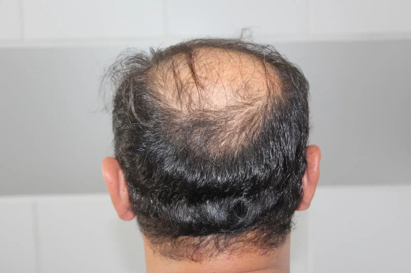
What Is the Donor Area in a Hair Transplant?
The donor area is the region from which hair follicles are collected for transplantation, usually across the back and sides of the scalp. Hair in these locations often shows greater resistance to the hormonal changes responsible for common pattern hair loss, making it a valuable source of transplantable follicles.
However, the donor area contains a limited number of follicular units. Hair transplantation redistributes existing follicles rather than creating new ones, so removing grafts permanently reduces the original supply.
The appearance of the donor area after surgery depends on how much hair remains, how evenly the grafts were extracted and how the surrounding scalp recovers.
Why Does the Donor Area Look Patchy After Surgery?
A patchy donor area may reflect several changes occurring at the same time. Immediately after surgery, shaving, small extraction wounds and temporary redness can make certain sections of the scalp appear thinner.
During the following weeks, some unharvested hairs may shed temporarily in response to surgical trauma. This process can create new-looking gaps even when the initial healing appeared satisfactory.
Another possibility is uneven graft removal. When too many follicles are extracted from a limited region, the remaining hair may no longer conceal the scalp adequately.
The most important distinction is whether the visible patches contain temporarily inactive follicles or areas where follicular units have been permanently removed.
How Does FUE Harvesting Change Donor Hair Density?
Follicular Unit Extraction, or FUE, involves removing individual follicular units with small surgical punches. Each extraction creates a tiny wound and transfers the selected follicle to another part of the scalp.
The surrounding follicles remain in place, but the original donor density decreases because the extracted units do not regenerate.
When extraction sites are appropriately distributed, the remaining hair can often conceal this reduction. Concentrating too many extractions in one location, however, may create visible gaps.
Why Does the Donor Area Look Uneven Immediately After FUE?
During the first days after FUE, the donor area may appear irregular because thousands of tiny extraction points are healing at slightly different rates.
Redness, pinpoint crusts and closely shaved hair can create a spotted pattern that makes the skin more visible than it normally would be.
The extraction sites usually close relatively quickly, and much of the superficial crusting clears within approximately one to two weeks.
As the surrounding hair grows longer, it begins covering the tiny healing marks, which can substantially improve the appearance.
However, surface healing should not be mistaken for the restoration of extracted follicles. The scalp can heal normally while its underlying follicular density remains permanently reduced.
What Is Donor Area Shock Loss?
Donor area shock loss is the temporary shedding of existing hairs near the regions affected by hair transplant surgery. It is also described as donor-site effluvium.
Surgical trauma, temporary disruption of local circulation and stress affecting nearby follicles may contribute to the shedding.
Unlike harvested grafts, these affected follicles remain in the donor area. If their growth structures are intact, they can resume producing hair as the scalp recovers.
Shock loss may produce localized bald-looking patches or more diffuse thinning across the harvested region.
Although the appearance can be alarming, temporary donor shedding is fundamentally different from permanent follicle removal or scarring.
What Are the Main Causes of Patchy Donor Hair?
Several mechanisms can create an uneven donor area, and their expected recovery differs considerably.
Temporary shock loss: Surgical stress causes some remaining hairs to shed, creating gaps that may improve when the affected follicles resume growth.
Donor overharvesting: Removing too many follicular units reduces permanent hair density, sometimes exposing the scalp despite normal wound healing.
Uneven extraction distribution: Concentrating graft removal in particular zones creates noticeable contrasts between heavily harvested and relatively untouched sections.
Scarring or postoperative inflammation: Extraction marks, folliculitis, infection or deeper tissue injury can affect the appearance and, in severe cases, damage additional follicles.
When Does Donor Shock Loss Begin?
Donor shock loss often becomes noticeable approximately two to four weeks after transplantation, although the timing is not identical in every case.
Someone may initially see the donor region recovering normally and then notice a new patch of thinning several weeks later. This delayed appearance is consistent with a temporary disturbance of the hair growth cycle.
Shedding can develop in sections adjacent to dense extraction sites or appear more diffusely across the back of the scalp.
However, timing alone cannot establish the diagnosis. Immediate gaps caused by permanent follicle removal and later shedding may coexist, which is why the pattern and progression of thinning also matter.
How Long Does Donor Shock Loss Last?
Temporary donor-site effluvium commonly improves over several months, with recovery often observed within approximately three to six months.
The scalp may stop shedding before the hair looks noticeably fuller. New hairs need time to grow long enough to contribute meaningful coverage, especially when the surrounding hair has already grown considerably.
Some areas recover faster than others, producing temporary differences in density.
The expected improvement assumes that the follicles remain intact. When scarring, substantial overharvesting or an underlying scalp disorder is present, waiting alone may not restore the missing coverage.
Persistent or worsening gaps deserve assessment rather than an assumption that all donor thinning represents normal shock loss.
What Is Donor Overharvesting?
Donor overharvesting occurs when too many follicular units are removed from a region relative to its available density and capacity to maintain natural coverage.
This can happen during a single extensive procedure or through several sessions that repeatedly use the same sections of scalp.
The result may be a permanently thin, irregular or visibly depleted donor area, sometimes described as a moth-eaten appearance.
The number of grafts extracted is only part of the calculation. Hair thickness, follicular density, scalp size and existing hair loss also determine how much removal can be concealed.
A high graft count does not automatically represent a better transplant when obtaining those grafts leaves the donor region visibly damaged.
Can Uneven Graft Extraction Create a Moth-Eaten Appearance?
Uneven extraction can leave alternating zones of relatively dense and sparse hair, creating the appearance of small, irregular bald patches.
These patches become particularly noticeable when many follicles are taken from one accessible section while neighboring areas retain most of their original hair.
Even if each extraction wound heals successfully, the overall pattern can remain visible because the missing follicular units do not regenerate.
The effect is sometimes called a window pattern when a clearly defined area shows substantially less coverage than the surrounding scalp.
Preventing this appearance requires balanced extraction across a suitable donor zone rather than concentrating removal wherever grafts are easiest to obtain.
Do Extracted Donor Hair Follicles Grow Back?
Follicular units completely removed during FUE do not grow back at their original donor sites. The harvested follicles have been transferred to the recipient area, leaving fewer hair-producing structures behind.
This biological limitation explains why donor management is central to hair transplantation.
However, the absence of a harvested follicle should not be confused with temporary shedding from an adjacent follicle that remains in the scalp.
Surrounding hairs affected by shock loss may regrow, while completely removed follicular units cannot.
Consequently, a donor area can gain visible density during recovery without ever returning to precisely the same number of follicles it contained before surgery.
How Do FUE Scars Affect Donor Area Appearance?
FUE leaves numerous small circular scars where follicular units were removed. These marks are often subtle once healing is complete and the surrounding hair provides sufficient coverage.
Some scars may appear lighter than the surrounding skin, creating small pale dots that become especially noticeable after a very short haircut.
Punch size, extraction spacing and wound healing influence how visible these marks become.
Although individual scars may be tiny, closely grouped extraction sites can collectively produce an uneven scalp appearance.
Hair growth around the scars may improve concealment, but ordinary skincare products cannot recreate follicles removed during extraction or completely erase established surgical scars.
Can FUT Cause Patchy Hair Around the Donor Scar?
Follicular Unit Transplantation, or FUT, involves removing a strip of hair-bearing scalp and closing the resulting wound.
Unlike FUE, which creates numerous small extraction marks, FUT usually leaves a linear scar across the donor region.
Some people experience temporary shedding near this incision because nearby follicles respond to surgical stress.
Excessive wound tension, wider scarring or impaired healing may also reduce coverage along the scar.
The distinction matters because thinning around a FUT incision may have a different anatomical cause from irregular patches after FUE.
Both methods can affect donor appearance, although they produce different patterns of scarring and hair loss.
Does Hair Length Make Donor Patchiness More Visible?
Hair length strongly influences how noticeable a depleted donor area appears.
When the back and sides of the head are shaved very closely, even moderate differences in follicular density can expose more scalp.
As the remaining hair grows longer, individual strands overlap and provide additional coverage, often making extraction marks and mild thinning less apparent.
Hair thickness and contrast between hair color and skin tone also affect visibility. Fine, dark hair against a light scalp may reveal gaps more readily than thicker hair with less color contrast.
However, camouflage through longer hair does not mean the missing follicles have returned.
A donor area that appears balanced with longer hair may still show permanent depletion when shaved.
Can Folliculitis or Infection Cause Donor Area Bald Spots?
Folliculitis can develop when hair follicles become inflamed after surgery, sometimes producing red bumps, pustules, tenderness or crusting.
Mild inflammation may resolve without permanent hair loss. More severe infection or repeated injury can damage surrounding tissue and delay healing.
Persistent redness, painful swelling and discharge should not be attributed to ordinary extraction wounds without examination.
Other inflammatory scalp conditions may also affect donor hair density independently of transplantation.
When bald patches appear alongside recurring pustules or significant tenderness, identifying and treating the underlying process becomes more important than using cosmetic hair growth products.
Can Poor Blood Supply Cause Permanent Donor Hair Loss?
Healthy blood circulation supports wound healing and the survival of hair follicles that remain in the donor region.
Excessive tissue trauma, closely concentrated extraction wounds or significant vascular injury can compromise the local healing environment.
In rare cases, serious circulation problems lead to skin necrosis, where affected tissue becomes damaged and may eventually scar.
Follicles destroyed by this process cannot resume normal growth.
Poor circulation can also contribute to temporary shedding without causing permanent tissue death, so these conditions should not be confused.
Severe pain, darkening skin or wounds that fail to heal require prompt medical attention because delayed treatment can increase the risk of lasting damage.
How Is Shock Loss Different From Permanent Donor Depletion?
The central difference is whether functioning hair follicles remain beneath the thinning area.
Shock loss affects hairs whose follicles are still present, allowing regrowth as the scalp recovers. Permanent depletion occurs when follicular units have been removed or destroyed.
The two conditions can initially look similar, especially when the donor area is closely shaved.
However, shock loss usually shows progressive improvement over subsequent months, while significantly overharvested regions may remain visibly sparse after normal healing.
These processes can also occur together, meaning a partially depleted donor area may improve somewhat without returning to its previous density.
A thorough assessment should consider both possibilities rather than treating every patch as temporary.
Which Warning Signs Require Medical Assessment?
Patchiness deserves particular attention when the appearance is accompanied by symptoms or changes suggesting something beyond uncomplicated healing.
Increasing pain or spreading redness: Worsening tenderness, warmth and expanding inflammation may indicate infection or another postoperative complication.
Pus, foul odor or persistent open wounds: Discharge and wounds that do not heal normally require evaluation rather than continued home treatment.
Dark or damaged-looking skin: Dusky discoloration, black crusts or tissue breakdown may indicate compromised circulation and require urgent assessment.
Progressive or persistent bald patches: Expanding gaps, obvious scarring or a lack of improvement over the expected recovery period may indicate permanent depletion or another hair-loss condition.
How Can Trichoscopy Help Diagnose Donor Area Thinning?
Trichoscopy uses magnification to examine the scalp, individual hairs and follicular openings without requiring a surgical procedure.
It can help assess differences in hair density, hair shaft thickness, extraction scars and signs of inflammation.
In suspected shock loss, surviving follicular structures may support the possibility of regrowth.
By contrast, scarred areas with absent follicular openings raise concern for permanent follicle destruction.
Trichoscopy may also identify miniaturization associated with an underlying hair-loss disorder, which can complicate the donor region's appearance.
When the cause remains uncertain, serial photographs or additional investigations may help document whether density is recovering, remaining stable or continuing to decline.
When Should Donor Area Density Start Improving?
During the first one to two weeks, improvement mainly involves healing of the extraction points and reduction of crusting.
Patchiness caused by short hair or visible wound marks may become less noticeable as the surrounding hair grows longer.
Temporary donor shock loss often appears after this initial stage, sometimes making the area look worse before it improves.
Over the following three to six months, recovering follicles may produce enough hair to improve visible density.
However, permanent extraction-related gaps do not fill with newly generated follicles.
If the donor region remains markedly sparse after several months, or if thinning continues to spread, further assessment can distinguish delayed regrowth from a more persistent problem.
Can Minoxidil Help Hair Regrow in the Donor Area?
Minoxidil may be considered when donor thinning involves follicles that remain capable of producing hair.
Some medical literature describes its use in supporting regrowth after donor-site shedding, although direct evidence for accelerating recovery in this specific situation remains limited.
Minoxidil cannot recreate follicular units removed during transplantation or restore hair growth in established scar tissue.
It can also irritate an injured scalp, particularly when applied before extraction sites have healed.
The presence of redness, pustules or open wounds should be evaluated before introducing a topical hair growth treatment.
The priority is to establish whether the problem involves temporary follicular inactivity, permanent depletion or an independent hair-loss condition.
How Should You Care for a Patchy Donor Area?
Appropriate aftercare helps protect healing follicles and skin, although it cannot replace grafts that were permanently removed.
Clean the scalp gently: Follow postoperative washing instructions, use appropriate products and avoid vigorous rubbing that could disturb healing extraction sites.
Leave crusts undisturbed: Scratching or picking can increase irritation, delay wound closure and potentially worsen scarring.
Reduce friction and irritation: Avoid tight headwear, harsh chemical products and very close shaving until the donor region has recovered sufficiently.
Monitor changes over time: Take photographs under consistent lighting and seek medical evaluation if patches expand, become painful or fail to improve.
Can an Overharvested Donor Area Be Repaired?
Repairing an overharvested donor area is more challenging than treating temporary shock loss because the missing follicular units do not regenerate.
In selected cases, remaining scalp follicles may be transplanted into visibly depleted regions. However, this approach requires an adequate supply of usable donor hair elsewhere.
Beard or body hair may offer additional grafts in certain situations, although differences in texture, growth behavior and appearance can limit their suitability.
Extensive scarring or unstable scalp inflammation may further restrict corrective options.
Can Scalp Micropigmentation Hide Donor Area Patchiness?
Scalp micropigmentation is a cosmetic technique that places small pigment deposits in the skin to simulate the appearance of short hair follicles.
It may reduce the visual contrast between sparse donor sections and the surrounding scalp, particularly when the hair is worn relatively short.
The technique can also help camouflage selected FUE scars or a linear FUT scar.
However, micropigmentation does not create hair, increase follicular density or reverse scarring.
The final appearance depends on pigment placement, color matching and the relationship between the treated skin and remaining hair.
It should be considered only after the scalp has healed and the underlying cause of patchiness has been adequately evaluated.
How Can Patchy Donor Areas Be Prevented?
Prevention begins with a realistic assessment of the donor area's follicular density, hair thickness, safe harvesting boundaries and long-term hair-loss pattern.
Graft extraction should be distributed evenly enough to preserve the visual coverage of the remaining hair.
Concentrating removal within a small section can create noticeable depletion even when the overall graft count seems reasonable.
Previous extraction sites must also be considered before another procedure, because repeated harvesting from the same zones progressively reduces available density.
The safest plan does not necessarily involve obtaining the highest possible number of grafts in one session.
Preserving enough donor hair for natural coverage and potential future procedures is an essential part of successful transplantation.
Is Another Hair Transplant Safe After Donor Area Thinning?
A second hair transplant may still be possible when enough stable donor hair remains, but visible thinning requires careful investigation before further graft removal.
If the existing patches result from temporary shock loss, recovery should be assessed before making decisions about additional harvesting.
When the donor area is already significantly depleted, extracting more scalp follicles may worsen the uneven appearance.
Previous surgery, miniaturization, remaining follicular density and possible scarring all influence whether another procedure is reasonable.
Alternative donor sources may be considered in selected cases, but they do not eliminate the limits imposed by the available hair supply.
Further surgery should not be used to solve one density problem by creating another.
Will a Patchy Donor Area Return to Normal?
A patchy donor area can improve considerably when temporary shock loss, short hair or early healing changes are responsible for the appearance.
As extraction wounds heal and surviving hairs grow longer, the scalp often looks more evenly covered. Temporary donor-site effluvium may also recover over several months when the underlying follicles remain healthy.
However, follicles removed during FUE do not regenerate in the donor region. Overharvesting, significant scarring or severe tissue injury can therefore leave permanent gaps.
The likelihood of recovery depends on identifying which of these processes is causing the visible thinning.
The most effective approach combines appropriate postoperative care with careful monitoring and timely investigation of persistent or worsening patches. Protecting the remaining donor hair is just as important as achieving good growth in the transplanted area, because both regions contribute to the overall result of hair restoration surgery.